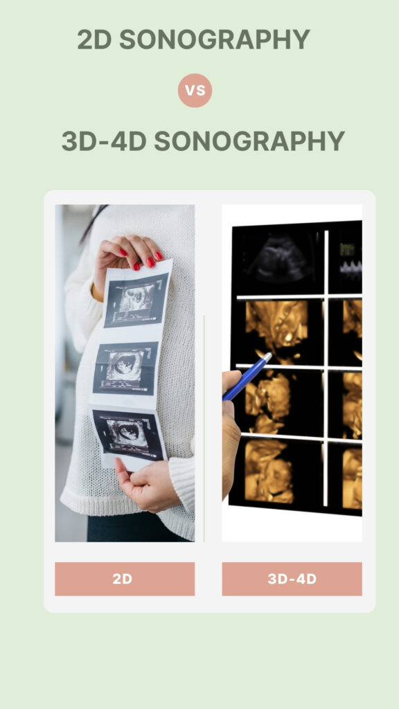
3D-4D Sonography

The use of 3D and 4D Sonography in Grace Women’s Hospital has been one of the significant advancements in prenatal imaging that provides expectant parents with a unique detailed outlook of their fetus. Below is a detailed description of how these technologies are used in this institution.
3D Sonography: Unlike conventional ultrasound, which presents flat images of the fetus, 3D Sonography at Grace Women’s Hospital creates three-dimensional images that offer a more realistic perspective on baby’s exterior features. The technology captures high-resolution pictures revealing unique facial details and physical appearance, thus helping doctors better evaluate the fetus’s growth status and detect abnormality in structures.
4D Sonography: On top of what it does, 4D usually shows movement or a live video feature. This way, the mother can see her child moving while still inside her womb. It enables one to observe activities such as smiling, yawning, and other movements, which bring about emotional bonding between parents and unborn children. It makes medical professionals much more capable of assessing the health and behavior patterns of the fetus.
Usage and Benefits: These advanced sonographic technologies are employed at Grace Women’s Hospital during routine prenatal care visits or when there is a need for extensive examination due to possible complications. Obstetricians and other healthcare providers can make more accurate diagnoses by seeing detailed images and movements, hence planning treatment when required. Moreover, it greatly improves expectant parents’ prenatal experiences.
Safety and Comfort: Both 3D and 4D ultrasounds are safe, just like traditional ultrasounds, since they use sound waves to make their pictures instead of radiation, therefore not harmful to either mothers or fetuses. Throughout these sessions, women expecting babies feel comfortable because they take place within an environment designed specially for them so that they can enjoy themselves through the fight against pregnancy stressors.
Conclusion: The usage of 3D and 4D sonography technologies by Grace Women’s Hospital provides a clear indication of its commitment to providing high-quality medical care with a human touch. Therefore, these advanced imaging techniques improve the quality of health service delivery and foster stronger bonds between parents and their babies yet to be born.
Difference Between Traditional and 3D-4D Sonography

1. Dimensional Depth and Detail:
–2D Sonography: Provides two-dimensional, flat fetus images. In most cases, these pictures are black-and-white views that present profiles or cross-sections helpful for medical evaluations but may be difficult for laypeople to interpret.
–3D Sonography: Produces three-dimensional images that allow us to see shapes, surfaces, or contours on the baby’s body. This provides more realistic external traits such as face, limbs, or organs, giving parents an image of what their child will look like and enabling doctors to get better anatomical details.
–4D Sonography: Adjoins time dimensionality into three-dimensional imagery, making it akin to live video footage showing fetal movements. It lets both parents and healthcare practitioners view actions like kicking, facial expressions, or even respiration
2. Diagnostic Value:
– 2D Sonography: Remains the standard for most diagnostic purposes, including confirming pregnancy and checking the fetal heartbeat. It is also used to examine fetal growth patterns, find out if there is any placental abnormality, and look for other abnormalities.
–3D and 4D Sonography: This provides improved vantage points that are useful, especially in the diagnosis of some physical anomalies such as cleft lip, spinal cord problems, or skeletal deformations that may be difficult to recognize on 2D scans. These also help in advanced bonding and psycho-preparation for parents having an abnormal fetus.
3. Patient Experience:
– 2D Sonography: However, it might not be as emotionally captivating to the parents because of the images’ low detailedness and a more scientific perspective.
– 3D and 4D Sonography: This has a more emotional touch due to the life-like features and movements of the baby within it; this further strengthens the relationship between unborn babies and their parents.
4. Usage Frequency:
– 2D Sonography: This is predominantly used for routine check-ups during pregnancy due to its effectiveness, simplicity, and affordability.
– 3D and 4D Sonography: They are commonly used selectively, mostly when there is a need for better imaging or when health care providers or anticipated parents would like a much clearer view of the fetus for personal or medical reasons.
5. Cost and Availability:
– 2D Sonography: More accessible than the advanced versions of this technology and generally costs less in most places.
– 3D and 4D Sonography: The cost implications make them expensive, with some facilities lacking them altogether, mainly due to higher acquisition costs of machines and more complex interpretation required.
